
The purpose of the study was to give a better understanding of the nature of the forehead bone in order to determine the best possible surgical or non-surgical treatments.
Abstract:
The study examined age-related and gender-related changes in the frontal bone and forehead. Cranial computed tomographic (CT) images from 157 Caucasian individuals were analyzed. The thickness and distance measurements of the frontal bone were taken to investigate age and gender differences. The results showed that the size of the male forehead decreases with age, and there were no significant differences compared to the female forehead in old age. The thickness of the frontal bone increased slightly in the lower forehead with age for both genders. However, in the upper forehead, the thickness of the frontal bone decreased significantly in males but showed no significant change in females.
Methods:
The study analyzed CT images obtained from 157 Caucasian individuals, including males and females from various age groups. Distance and thickness measurements of the frontal bone were performed using multi-planar computed tomography imaging. The measurements were taken at different angles and locations on the forehead.
Results:
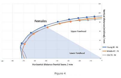
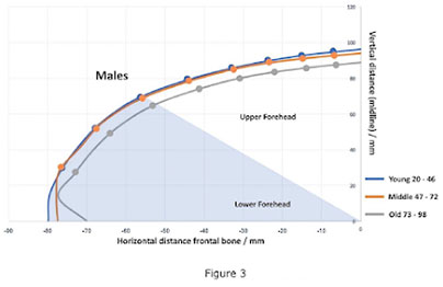
Females had smaller mean distance measurements compared to males in both young and middle-aged groups. However, in the old age group, there was no significant difference between genders as males reduced overall in mean distances. The distance between the posterior clinoid process and the internal lamella of the frontal bone decreased with age in males. The thickness of the frontal bone in the lower forehead increased slightly with age for both genders. In the upper forehead, the thickness of the frontal bone decreased significantly with age in males but showed no significant change in females.
Conclusion:
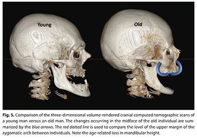
The study found that the shape of the frontal bone varies in young individuals of different genders and undergoes complex changes with age. Understanding these changes in both the bone and soft tissues of the forehead is important for determining the best surgical and nonsurgical treatment options. These findings contribute significantly to our knowledge of age-related changes in the frontal bone, particularly in a Caucasian population.